 This is the R1 ccPCR for my target protein H1N1 Hemagglutinin and I have found nothing amplified in the NTC. And as a side note, the 100 bp ladders turned out to be very faint and I'm not the only person whose had really faint ladders.
This space represents the ideas, views, opinions, projects and data of researchers within the Aptamer Stream of the Freshman Research Initiative, a program developed at the University of Texas at Austin. These are projects we currently have in the pipeline.
- We're not exclusive...we are SELECTIVE!
Labels
- Projects (81)
- Targets (75)
- Informational (48)
- Data (24)
- Opportunities (24)
- FAQ (16)
- Target Proposal (15)
- Guidelines (12)
- projects targets (9)
- Journal Articles (7)
- final manuscript (4)
- Abstract (3)
- Help (3)
- artifact (2)
- band (2)
- Dustin Taylor final manuscript (1)
- FGF-2 (1)
- FGF-b (1)
- Fall 2012 (1)
- Juan Herrejon CCR5 Progress Report (1)
- Phttp://www.blogger.com/img/blank.gifrojects (1)
No artifact in the N50 pool.
 This is the R1 ccPCR for my target protein H1N1 Hemagglutinin and I have found nothing amplified in the NTC. And as a side note, the 100 bp ladders turned out to be very faint and I'm not the only person whose had really faint ladders.
HIV-1 Gag p24 First Progress Report--Camille Alilaen
October 18, 2011
Fall 2011
Pool N34, HIV-1 Gag p24
R50 Selection Against CD105/Endoglin - Progress Report #1 (10/18/11)


KRAS R1 Progress Report - Ryan Lannan
October 18, 2011
N58, RNA, KRAS
Progress Report 1
Background information on and justification for a KRAS selection can be found here as an abstract and proposal. The KRAS selection will be performed using the “toggle” technique. This technique involves the use of two different selection methods in order to remove background binders to the different binding mediums. Due to KRAS’s size (50 kDa) a filter-based selection is possible. And as for the other selection method, the His tag attached to KRAS allows for a bead-based selection using nickel beads. The selection will alternate between filter and bead-based selections, starting with a filter selection. The first round of filter-based selection was performed against KRAS with the following conditions:
Round One Conditions
Nucleic Acid/Protein Ratio: 400-pmol/400-pmol
Buffer: 1X PBS SELEX with MgCl2 salts (pH 7.4)
Incubation Temperature/Time: 37 degrees C for 40 minutes
Wash Frequency and Volume: Three 100 uL washes
Progress, Results and Discussion
To begin the selection, an RNA binding reaction was prepared with 400 pmol of N58 R0 pool diluted by 1X PBS SELEX buffer. The reaction was then denatured by heating it to 65 degrees C for five minutes. 400 pmol of KRAS was added to the reaction and incubated for 40 minutes at 37 degrees C to allow both nucleic acids and protein to reach a stable equilibrium at physiological temperatures. The nitrocellulose filter was set up and washed three times using 1X PBS SELEX buffer. The 100 uL binding reaction created earlier was then added to the filter and the liquid was pushed through using a syringe, hopefully leaving behind the target and any possible binders. Three 100 uL washes with 1X PBS SELEX buffer were then performed to remove any non-binders. After washing, an elution was performed on the filter using 200 uL Elution buffer (5M urea and 25mM EDTA). The elution reaction was then heated at 90-100 degrees C for five minutes. The elution buffer was then removed and another 200 uL of elution buffer was reapplied and the reaction was reheated. The combined 400 uL was then ran through ethanol precipitation to concentrate the nucleic acids. The pellet produced from ethanol precipitation was resuspended in 10 uL diH2O and an initial reverse transcription solution was performed with 7 uL of the RNA solution, 20 uM of N58 reverse primer and 50 uM dNTPs. This initial mix was incubated for five minutes at 65 degrees C, and allowed to cool. 1X First Strand Buffer, 10 mM Dtt and 1 uL SSII reverse transcriptase were added and the reaction was run using these steps: 42 degrees C for 50 minutes, 70 degrees C for 15 minutes and a cooling step.
To amplify the newly created ssDNA, a PCR reaction had to be performed. In order to determine the optimal number of cycles and troubleshoot possible selection problems, cycle course PCR was run before large-scale PCR. The following reaction was created for E1, 1X PCR buffer, 0.2 mM dNTP, 0.4 uM forward and reverse N58 primer, 2 uL ssDNA, and 0.8 uL Taq DNA polymerase. 5 uL was removed from this PCR reaction at each chosen cycle, and another aliquot was removed from a cycle without template at 18 cycles as a no-template control which came out negative (see Figure 1 below). An agarose gel was run with these 5 uL aliquots (Figure 1).
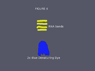
Looking at band intensity, it was determined that 12 was the optimal number of cycles for lsPCR (in a filter selection there are no other washes, cycle course gels on even rounds will be more informative as they utilize beads and have multiple washes). Large-scale PCR was ran on the remainder of the ssDNA, creating 6 PCR reactions identical to the one described for cycle course. After lsPCR, an ethanol precipitation was performed. The pellet created was resuspended in 20 uL of diH2O. A transcription reaction was then prepared with the following composition: 1X Ampliscribe Transcription Buffer, 10 mM DTT, 7.5 mM of each NTP, 5 uL of ds DNA from lsPCR and 2 uL of T7 enzyme. The reaction was incubated overnight at 37 degrees C. After transcription, a PAGE gel (represented very roughly by Figure 2) was ran to purify the RNA from the transcription components, which was successful (showing a distinct band). Ethanol precipitation was then performed, and the pellet resuspended in 30 uL 1X PBS SELEX buffer.
Figure 2 – KRAS R1, N58
Problems Encountered
During filter selection at the beginning of the round, a hissing noise was noted while washing the filter. This could indicate that a small tear had formed in the filter, causing liquid to flow through without passing through the filter. Seeing as the binding reaction had already been applied, I continued onwards, hoping that the tear was small enough to not cause a significant loss of my binding reaction.
Additional Work
Besides small tasks I have performed such as re-aliquotting protein and glycogen, or processing protein deliveries, I have started helping Oliver with his closed-MBP selection. He is currently cloning his rounds to determine sequences, and I have started to help him by regenerating his R9, R11 and R12 pools to dsDNA.
Conclusion and Future Work
I will continue to help Oliver with his selection, and will soon perform cloning on his (now) dsDNA pools. The concentration of my R1 gold was 1768.4 ng/uL. This is a good number quantity of RNA, but I am hoping to see a decrease in concentration next round indicating that my switching of selection methods has significantly decreased background binders. R2 has already been started with Nickel beads, and I hope to finish the round within the week and start the cloning procedure for Oliver’s selection.


