Showing posts with label Projects. Show all posts

Nucleic Acid Aptamer Selection Against Angiotensin II


Nucleic Acid Aptamer Selection Against Angiotensin II
Owais Jamil
Nucleic Acid Pool: N50 RNA Pool
Target: Angiotensin II


Hypertension or more commonly referred to as high blood pressure, is a condition in which the blood pressure in the arteries increases, increasing a person’s risk of heart disease. This includes life threatening ailments such as stroke or heart failure (1). According to the Center for Disease Control, cardiovascular disease is the leading cause of death in the United States, affecting over two million each year. Deterrence of hypertension by means of would eliminate the risk of patients experiencing subsequent cardiovascular events and preventing long term damage to vital organs or death.

One of the causes of hypertension is an abnormality in the renin-angiotensin- aldosterone system (RAAS), a hormone system that regulates cardiac function by controlling blood pressure (2). Angiotensin II is a peptide hormone that is a part of this system that stimulates the release of aldosterone, a steroid hormone that causes blood pressure to increase by narrowing blood vessels. A patient suffering from hypertension would have an overly active RAAS and large quantities of aldosterone, and angiotensin II in their bloodstream. Inhibition of angiotensin II could prevent unsafe rises in blood pressure, thus preventing further complications in a patient’s condition.

A treatment for hypertension would be to use an RNA aptamer with a high specificity for angiotensin II. An aptamer is a short strand of oligonucleotides that has a high binding affinity for a specific macromolecular target (3). Aptamers can be used for a variety of functions, one of which includes inhibiting the function of its target. Thus, an aptamer could be an effective treatment by inhibiting the function of angiotensin II.

Specific Aim 1: Perform the Systematic Evolution of Ligands by Exponential Enrichment (SELEX) method to select an RNA aptamer against angiotensin II. 

Specific Aim 2: Modify aptamer for inhibition of angiotensin II. After selecting for an aptamer with a high binding affinity for angiotensin II, it can be modified for use as an inhibitor, as angiotensin II is very abundant in patients with high blood pressure. This would prevent the release of aldosterone, the hormone subsequently leading to a drop in blood pressure (4).


Figure 1 Specific Aim 2. By using an aptamer to inhibit the function of angiotensin II, aldosterone will not be released and blood pressure can be stabilized. 


Biotinylated Angiotensin II (MW = 1.3kDa) can be ordered from AnaSpec.
Catalog Number: 60276-1
Cost for 1mg: $66
Cost per round:  $0.01

References

1. Constantino, I., Gorelick P. B. 2003 “Hypertension, Angiotensin, and Stoke: Beyond Blood Pressure” Stroke (35). 348-350
2. Peach, M. 1977 “Renin-Angiotensin System: Biochemistry and Mechanisms of Action” Physiological Review (57). 313-370
3. Elligton, A.D., Szostak J.W. 1990 “In vitro selection of RNA molecules that bind to specific ligands” Nature (346). 818-822
4. Crowley, S.D., Gurley, S.B., Herrera, M. J., Ruiz, P., Griffiths, R., Kumar, A.P., Hyung-Suk, K., Smithies, O., Le, T. H., Coffman, T. M. 2006 “Angiotensin II causes hypertension and cardiac hypertrophy through its receptors in the kidney” PNAS (103). 17985-17990


Click here to view full target proposal.
(http://dl.dropbox.com/u/106599935/Owais%20Jamil%20Target%20Proposal.pdf)

Click here to view the first progress report.
(http://dl.dropbox.com/u/106599935/Owais%20Jamil%20Progress%20Report%201.pdf)

Click here to view the second progress report.
(http://dl.dropbox.com/u/106599935/Owais%20Jamil%20Progress%20Report%202.pdf)

Click here to view the final manuscript
(http://dl.dropbox.com/u/106599935/Owais%20Jamil%20Final%20Manuscript%20Fall%202012.pdf)

Nucleic Acid Aptamer Selection Against FasR for Inhibition of Nerve Cell Apoptosis


Owais Jamil
Fall 2012
September 4, 2012
N50 RNA Pool, FasR/CD95

Nucleic Acid Aptamer Selection Against FasR for Inhibition of Nerve Cell Apoptosis

Abstract
Permanent damage to the nervous system can be caused by severe physical trauma to the peripheral nervous system. Following this trauma, one of the ways loss of function occurs is secondary damage to the central nervous system, which occurs in the form of neuron death. If this can be prevented, victims will be able to retain motor and cognitive ability (2).

The Fas Receptor (FasR, also known as CD95) is a member of the tumor necrosis factor protein family that found on the surface of somatic cells and functions in cell death. When the Fas ligand (FasL) binds to FasR, a death inducing signaling complex (DISC) is formed (1). This triggers a downstream signaling cascade ending with apoptosis. In nervous system peripherals that have experienced trauma, FasR and FasL have been shown to be present in high quantities (4).

Apoptosis is the process of programmed cell death triggered by various signals or inducing factors. In the event of a spinal cord injury, damage occurs at the cellular level as a result of apoptosis. In healthy nervous systems, apoptosis pathways are used to eliminate unused neuronal connections and eliminate damaged cells (3). However, in the case of severe trauma, cell death can become uncontrolled, eliminating not only damaged cells but also healthy cells (2). These damages are irreversible and can lead to severe loss of nervous system function, and even death.

Specific Aim:
In the nervous system, when FasL binds to FasR, the DISC begins the pathway within the cell to lead to its death. To prevent this, perform the SELEX method to select an RNA aptamer with a high binding affinity for FasR. This aptamer would serve as a means to inhibit the function of FasR in healthy neurons that surround damaged nervous tissue, in hopes of preventing increased damage to the irreparable nerve tissue.


Figure 1. By inhibiting the function of FasR in healthy neurons surrounding trauma, loss of function can be minimized.

FasR/CD95 can be purchased from BD Pharmingen (Catalog#554256) at the price of $255 for 500 micrograms. (http://www.bdbiosciences.com/ptProduct.jsp?prodId=10379&catyId=745875)

References:

1. Kim, J.W., Choi, E., O Joe, C. 2000 “Activation of death-inducing signaling complex (DISC) by pro-apoptotic C-terminal fragment of RIP” Oncogene (19). 4491-4499

2. Beattie, M.S. 2004 “Inflammation and apoptosis: linked therapeutic targets in spinal cord injury” Trends in Molecular Medicine (12). 580-583

3. Groene, H-J., Herr, I., Krammer, P.H., Martin-Villalba, A. 2006 “Control of neuronal brancing by the death receptor CD95 (Fas/Apo-1)” Cell Death and Differentiation (13). 31-40

4. Shohami, E., Trentz, O., Kossman, T., Morganti-Kossman, M.C. 2007 “Immunohistochemical characterization of Fas (CD95) and Fas Ligand (FasL/CD95L) expression in injured brain: Relationship with neuronal cell death and inflammatory mediators” Histol Histopathol (22) 235-250

Nucleic Acid Aptamer Selection against Endoglin (CD105) for the Inhibition of Tumor Cells


Vicki Oladoyin

September 18, 2012

FRI: Aptamer

N40B Pool, RNA, CD105

Nucleic Acid Aptamer Selection against Endoglin (CD105) for the Inhibition of Tumors

Abstract:

           Endoglin (CD105) is a cell membrane glycoprotein expressed on cellular lineages within the vascular system and is involved in blood vessel development (Fonsatti 2003). It has been discovered that endoglin may be involved with tumors associated with the vascular endothelium, because endoglin has been found to be over-expressed on proliferating endothelial cells of both peri-and intra-tumoral blood vessels (Fonsatti 2003).  For research purposes, an aptamer against mouse endoglin will be selected via in vitro SELEX (systematic evolution of ligands by exponential enrichment).  Furthermore, the practical implications of using an aptamer selected against endoglin to inhibit the formation of tumors will be discussed.

Specific Aim 1: Identifying an Aptamer Against Endoglin

Indentifying a high affinity and specific RNA aptamer against endoglin would be beneficial, because endoglin is expressed on over-proliferating endothelial cells as represented in Figure 1.  An aptamer selected against endoglin would therefore be useful for diagnosis and possibly the development of a therapeutic drug to inhibit the over-proliferation of tumor cells.

Specific Aim 2: Identifying an Anti-Endoglin Aptamer

            Identifying an anti-endoglin aptamer would be a useful tool for the studying of the functions and processes of the endoglin glycoprotein in the human body and its role in the formation of blood vessels.  This could then lead to a better understanding of angiogenesis, the physiological process involving the growth of new blood vessels from pre-existing vessels (Wahl 2004).

Endoglin (CD105) protein can be purchased from Sino Biological Inc. at a price of $290 per 100ug; its call number is 50407-M08H.  It has a calculated molecular mass of 61.2kDa.  The cost per round will be about $35.50. This amount will allow eight rounds of selection to be performed using 200 pmols of the target per round.  The company's phone number is 86-400-890-9989.


References

1.  Abdalla S., and M Letarte. "Hereditary Haemorrhagic Telangiectasia: Current Views on Genetics and Mechanisms of Disease."J Med Genet (2006): 97-110.


2.  Dallas, Nikolaos, Shaija Samuel, Ling Xia, Fan Fan, Michael Gray, Sherry Lim, and Lee Ellis. "Endoglin (CD105): a marker of tumor vasculature and potential target for therapy." Clinical cancer research : an official journal of the American Association for Cancer Research 14.7 (2008): 1931-1937.


3.  Ellis, L. (2008) “Endoglin (CD105): A Marker of Tumor Vasculature and Potential Target for Therapy” Clinical Cancer Research. 14:1931.


4.  Ester, F. (2003) “Endoglin (CD105): a powerful therapeutic target on tumor-associated angiogenetic blood vessels.” Oncogene. 22:6557-6563.


5.  Fonsatti E, Sigalotti L, Arslan P, Altomonte M, Maio M. Emerging role of endoglin (CD105) as a marker of angiogenesis with clinical potential in human malignancies. Curr Cancer Drug Targets. 2003 Dec;3(6):427-32.


6.  "HHT Foundation International: About HHT." HHT Foundation International: Hereditary Hemorrhagic Telangiectasia – Osler-Weber-Rendu. N.p., n.d. Web. 2 Sept. 2012.

7.  P. Shannon Pendergast, H. Nicholas March, Dilara Grate, Judith M. Healy, Martin Stanton. "Nucleic Acid Aptamers for Target Validation and Therapeutic Application." J Biomol Tech. 2005 September;16(3): 224-234. Arbl.cvmbs.colostate.edu.


8.  Siemann DW. "Vascular Targeting Agents". Horizons in Cancer Therapeutics: From Bench to Bedside. 2002;3(2):4-15.


9. Stoltenburg, R., C. Reinemann, and B. Strehlitz. "SELEX—A (r)evolutionary Method to   Generate High-affinity Nucleic Acid Ligands." Biomolecular Engineering 24.4 (2007): 381-403.


10. Wikström, P., Lissbrant, I. F., Stattin, P., Egevad, L. and Bergh, A. (2002), Endoglin (CD105) is expressed on immature blood vessels and is a marker for survival in prostate cancer. Prostate, 51: 268–275. doi: 10.1002/pros.10083.


No artifact in the N50 pool.

This is the R1 ccPCR for my target protein H1N1 Hemagglutinin and I have found nothing amplified in the NTC. And as a side note, the 100 bp ladders turned out to be very faint and I'm not the only person whose had really faint ladders.


HIV-1 Gag p24 First Progress Report--Camille Alilaen

Camille Alilaen
October 18, 2011
Fall 2011
Pool N34, HIV-1 Gag p24


Abstract and Proposal Link: http://aptamerstream.blogspot.com/2011/08/aptamer-selection-against-hiv-1-gag-p24.html

Progress, Results and Discussion:
            In the week following the completion of the second practice round, the RNA selection process began against the HIV-1 Gag p24 protein. The conditions selected were tailored for the protein itself, and will become more restricted in order to select for the best binding aptamers. Because the protein was tagged with His, nickel-NTA beads were used to incubate the RNA with the protein. This incubation lasted for 25 minutes and was kept at 37*C for its duration, in order to mimic body temperature. Three washes using one volume (100 uL) was used to wash the loose or non-binding RNA from the beads, and the buffer used was Tris at pH 7.3.
            After ethanol precipitation and reverse transcription were performed on the reaction, cycle course was done on the resulting DNA. After withdrawing 5 uL from the E1, W3, and W0 reactions every 6th, 9th, 12th, 15th, and 20th cycle, ethidium bromide was added to each of the 15 tubes. The reactions run for 35 minutes in TBE buffer at 110 V in a 3.8% agarose gel. It was determined that the optimal cycle for running large scale PCR without overamplifying the DNA was 10 cycles, as seen on Figure 1 below.


 Figure 1: PCR gel for R1. The 3.8% agarose gel was run at 110 V for about 35 minutes in TBE buffer. Samples were taken starting at cycle 6 until cycle 15 in 3 cycle increments, then at cycle 20.
            A no template control was made for the cycle course PCR, however, it was not added in with the cycle course samples due to human error. Therefore, it was run the next day by itself in another 3.8% agarose gel for 30 minutes at 105 V in TBE buffer. No bands appeared, therefore, all reagents were free of contaminants (see Figure 2). Large scale PCR was also performed on 6 tubes of 100 uL of DNA for 10 cycles. These reactions were then ethanol precipitated, and transcription was performed on the result.


Figure 2: The no template control was run in a 3.8% agarose gel in TBE buffer at 105 V for 30 minutes alongside the ladder. No bands appeared, and the small dot that appeared was assumed to be caused by human error while taking the picture.

Problems Encountered:
            There were no problems with working with the protein during target immobilization and incubation, and binding and selection as well as ethanol precipitation and reverse transcription went smoothly. However, when the cycle course gel was run, it was noted that an unusual amount of bands appeared in W3. Although this result is not unheard of, it is still undesirable as it shows that there are many loose binders. In addition to this, during large scale PCR it was noticed that instead of 600 uL of reagents, there were about 500 uL after PCR. This change in volume could be due to evaporation or human error (i.e not adding a reagent, which could cause failure while running the PAGE gel).

Additional Work:
            When the lab used up all the 3.8% agarose gel, I helped Austin to prepare more, which was a great learning experience in case no mentors are present and the agarose is needed. I also helped him to aliquot 4 uM of dNTP’s when we ran out of stock.


Conclusion and Future Work:
            Since research on HIV-1 Gag p24 has begun, most of a round of selection has been completed. Cycle course was successful, even though there were more bands in W3 than desirable. The no template control was clear, and all that remains to complete is the PAGE gel and elution of RNA. Over the next weeks I plan to complete this round and about three or four more others, prior to the next progress report.


P.S Gwen sorry for posting so late!!

R50 Selection Against CD105/Endoglin - Progress Report #1 (10/18/11)


Progress, Results and Discussion

Dry protein CD105/Endoglin(1600pmol) was resuspended in PBS buffer to achieve desired working concentration of around 20uM. Aliquots consisted of 3X200pmol (10ul) and 10X100pmol (5ul). Binding reaction (100ul) was made with 400pmol R50 pool and 200pmol target protein. Filter-based selection was designated optimum partioning method for size of protein (61.2kDa).

Millipore nitrocellulose membrane filter (0.45um) was pre-washed with 100ul 1X PBS SELEX buffer and five total washes executed after addition of binding reaction. RNA was eluted with 400ul elution buffer (5M urea and 25mM EDTA buffer). Ethanol Precipitation of RNA was performed with 1/10th volume 3M NaOAc, 3ul glycoblue, 2.5 volumes 100% ethanol. Reverse transcription achieved with following conditions: incubate at 42°C for 50 minutes and heat inactivate the enzyme at 70°C for 15 minutes. The correct R50 T-7 Forward primer and R50 reverse primer were used to prepare the 100ul PCR reaction with 2ul ssDNA from reverse transcription which was performed on W0, W5, and E1. Reactions were cycled for 20 cycles under following conditions: initial at 94°C for 2 minutes, denature at 92°C for 45 seconds, anneal at 54°C for 45 seconds, elongate at 72°C for 60 seconds. Five ul aliquots were extracted during end of cycles as shown in Figure 1 in orange.


As seen in figure 1, 11 cycles was chosen to be performed during large scale PCR. Ethanol precipitation of PCR yielded dsDNA for subsequent transcription reaction. Five ul precipitated dsDNA was transcribed using the Ampliscribe Transcription Kit from Epicentre and incubated at 37°C for 16 hours. RNA was purified with 8% Polyacrylamide Gel Electrophoresis, 10% APS and TEMED. Denaturing bromophenol blue (2X) dye was used in a 1X TE TBE rig. Visualization of RNA shadow in gel through UV illuminator was very small and faint. Crush soak elution and precipitation was continued with the ssDNA PAGE band. Nanodrop specing revealed 78ng/ul or a concentration of 2.33uM. This concentration was too low to continue on to the next round and steps to address this issue are discussed in following section.


Problems Encountered

Initially, 400pmol of target protein was to be added to 400pmol (20ul) pool to begin selection. However the pool mixture RNA was not denatured before protein was added and the process had to be repeated. With only ~1400pmol left of protein, 200pmol (10ul) was used in consideration of minimal remaining amount. While adding protein (W0), no resistance against filter was observed unlike in pre-wash. The filter was then assumed to contain a hole, but selection was continued and stringency decrease noted. A no-template negative control failed to be performed, but will be in future gels.

Concentration of RNA after one round deemed insufficient to proceed. Possible reasons for low amounts were mainly attributed to small RNA shadow observed in PAGE. Failure during transcription posed as leading cause for low detection. Therefore an agarose gel was run to validate this hypothesis as seen in figure 2.


As seen in Figure 2, the dsDNA band was found to be around equal length to the 100bp. Therefore the problem occurred after large scale PCR and transcription needed to be duplicated with DNA from PCR. Also, the smudging around what should be a clearly defined band was noted and could indicate possible over amplification. Transcription was then repeated with careful precaution to use of reagents and alteration of Ampliscribe Kit then that of the previous used. It was then realized that 0.1M DTT had been used instead of 100mM thus explaining reason for low amplification during first transcription reaction. This new transcription reaction that used 100mM DTT was incubated for 16 hours. When placed in incubator no precipitate or evidence of a nucleotide crashing out was evident however when retrieved the next morning clear evidence of white precipitant was observed. DNA template was still removed and preparation of PAGE in hopes of a still successful transcription reaction.


Conclusion and Future Work

As of October 18th, 2011 one round of selection has been executed using the R50 pool against Endoglin/CD105. However, this round was not completely successful due to insufficient RNA concentration amount of 2.33uM. Error in amount of DTT was concluded to be reason for transcription failure and correct amounts were used in new reaction. PAGE on second transcription reaction will hopefully reveal successful concentrations of RNA to continue on to second round where a negative selection will also be performed. In the next reporting period I hope to accomplish a couple more rounds of selection.


Final Manuscript: https://docs.google.com/document/d/1DRo2m--Hr9V4lYq8MnboE8rvMq-Y5Hyni1zN0zEZ2GU/edit


KRAS R1 Progress Report - Ryan Lannan

Ryan Lannan

October 18, 2011

N58, RNA, KRAS

Progress Report 1

Background information on and justification for a KRAS selection can be found here as an abstract and proposal. The KRAS selection will be performed using the “toggle” technique. This technique involves the use of two different selection methods in order to remove background binders to the different binding mediums. Due to KRAS’s size (50 kDa) a filter-based selection is possible. And as for the other selection method, the His tag attached to KRAS allows for a bead-based selection using nickel beads. The selection will alternate between filter and bead-based selections, starting with a filter selection. The first round of filter-based selection was performed against KRAS with the following conditions:

Round One Conditions

Nucleic Acid/Protein Ratio: 400-pmol/400-pmol

Buffer: 1X PBS SELEX with MgCl2 salts (pH 7.4)

Incubation Temperature/Time: 37 degrees C for 40 minutes

Wash Frequency and Volume: Three 100 uL washes

Progress, Results and Discussion

To begin the selection, an RNA binding reaction was prepared with 400 pmol of N58 R0 pool diluted by 1X PBS SELEX buffer. The reaction was then denatured by heating it to 65 degrees C for five minutes. 400 pmol of KRAS was added to the reaction and incubated for 40 minutes at 37 degrees C to allow both nucleic acids and protein to reach a stable equilibrium at physiological temperatures. The nitrocellulose filter was set up and washed three times using 1X PBS SELEX buffer. The 100 uL binding reaction created earlier was then added to the filter and the liquid was pushed through using a syringe, hopefully leaving behind the target and any possible binders. Three 100 uL washes with 1X PBS SELEX buffer were then performed to remove any non-binders. After washing, an elution was performed on the filter using 200 uL Elution buffer (5M urea and 25mM EDTA). The elution reaction was then heated at 90-100 degrees C for five minutes. The elution buffer was then removed and another 200 uL of elution buffer was reapplied and the reaction was reheated. The combined 400 uL was then ran through ethanol precipitation to concentrate the nucleic acids. The pellet produced from ethanol precipitation was resuspended in 10 uL diH2O and an initial reverse transcription solution was performed with 7 uL of the RNA solution, 20 uM of N58 reverse primer and 50 uM dNTPs. This initial mix was incubated for five minutes at 65 degrees C, and allowed to cool. 1X First Strand Buffer, 10 mM Dtt and 1 uL SSII reverse transcriptase were added and the reaction was run using these steps: 42 degrees C for 50 minutes, 70 degrees C for 15 minutes and a cooling step.

To amplify the newly created ssDNA, a PCR reaction had to be performed. In order to determine the optimal number of cycles and troubleshoot possible selection problems, cycle course PCR was run before large-scale PCR. The following reaction was created for E1, 1X PCR buffer, 0.2 mM dNTP, 0.4 uM forward and reverse N58 primer, 2 uL ssDNA, and 0.8 uL Taq DNA polymerase. 5 uL was removed from this PCR reaction at each chosen cycle, and another aliquot was removed from a cycle without template at 18 cycles as a no-template control which came out negative (see Figure 1 below). An agarose gel was run with these 5 uL aliquots (Figure 1).


Looking at band intensity, it was determined that 12 was the optimal number of cycles for lsPCR (in a filter selection there are no other washes, cycle course gels on even rounds will be more informative as they utilize beads and have multiple washes). Large-scale PCR was ran on the remainder of the ssDNA, creating 6 PCR reactions identical to the one described for cycle course. After lsPCR, an ethanol precipitation was performed. The pellet created was resuspended in 20 uL of diH2O. A transcription reaction was then prepared with the following composition: 1X Ampliscribe Transcription Buffer, 10 mM DTT, 7.5 mM of each NTP, 5 uL of ds DNA from lsPCR and 2 uL of T7 enzyme. The reaction was incubated overnight at 37 degrees C. After transcription, a PAGE gel (represented very roughly by Figure 2) was ran to purify the RNA from the transcription components, which was successful (showing a distinct band). Ethanol precipitation was then performed, and the pellet resuspended in 30 uL 1X PBS SELEX buffer.

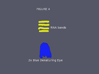
Figure 2 – KRAS R1, N58

Problems Encountered

During filter selection at the beginning of the round, a hissing noise was noted while washing the filter. This could indicate that a small tear had formed in the filter, causing liquid to flow through without passing through the filter. Seeing as the binding reaction had already been applied, I continued onwards, hoping that the tear was small enough to not cause a significant loss of my binding reaction.

Additional Work

Besides small tasks I have performed such as re-aliquotting protein and glycogen, or processing protein deliveries, I have started helping Oliver with his closed-MBP selection. He is currently cloning his rounds to determine sequences, and I have started to help him by regenerating his R9, R11 and R12 pools to dsDNA.

Conclusion and Future Work

I will continue to help Oliver with his selection, and will soon perform cloning on his (now) dsDNA pools. The concentration of my R1 gold was 1768.4 ng/uL. This is a good number quantity of RNA, but I am hoping to see a decrease in concentration next round indicating that my switching of selection methods has significantly decreased background binders. R2 has already been started with Nickel beads, and I hope to finish the round within the week and start the cloning procedure for Oliver’s selection.